Abstract Background: We studied the relationship between trismus (maximum interincisor opening [MIO] ≤35 mm) and the dose to the ipsilateral masseter muscle (iMM) and ipsilateral medial pterygoid muscle (iMPM). Methods: Pretreatment and post-treatment measurement of MIO at 13 weeks revealed 17% of trismus cases in 83 patients treated with chemoradiation and intensity-modulated radiation therapy. Logistic regression models were fitted with dose parameters of the iMM and iMPM and baseline MIO (bMIO). A risk classification tree was generated to obtain optimal cut-off values and risk groups. Results: Dose levels of iMM and iMPM were highly correlated due to proximity. Both iMPM and iMM dose parameters were predictive for trismus, especially mean dose and intermediate dose volume parameters. Adding bMIO, significantly improved Normal Tissue Complication Probability (NTCP) models. Optimal cutoffs were 58 Gy (mean dose iMPM), 22 Gy (mean dose iMM) and 46 mm (bMIO). Conclusions: Both iMPM and iMM doses, as well as bMIO, are clinically relevant parameters for trismus prediction.
DOCUMENT

Chest imaging plays a pivotal role in screening and monitoring patients, and various predictive artificial intelligence (AI) models have been developed in support of this. However, little is known about the effect of decreasing the radiation dose and, thus, image quality on AI performance. This study aims to design a low-dose simulation and evaluate the effect of this simulation on the performance of CNNs in plain chest radiography. Seven pathology labels and corresponding images from Medical Information Mart for Intensive Care datasets were used to train AI models at two spatial resolutions. These 14 models were tested using the original images, 50% and 75% low-dose simulations. We compared the area under the receiver operator characteristic (AUROC) of the original images and both simulations using DeLong testing. The average absolute change in AUROC related to simulated dose reduction for both resolutions was <0.005, and none exceeded a change of 0.014. Of the 28 test sets, 6 were significantly different. An assessment of predictions, performed through the splitting of the data by gender and patient positioning, showed a similar trend. The effect of simulated dose reductions on CNN performance, although significant in 6 of 28 cases, has minimal clinical impact. The effect of patient positioning exceeds that of dose reduction.
LINK
Objectives: Children have a greater risk from radiation, per unit dose, due to increased radiosensitivity and longer life expectancies. It is of paramount importance to reduce the radiation dose received by children.This research concerns chest CT examinations on paediatric patients. The purpose of this study was to compare the image quality and the dose received from imaging with images reconstructed with filtered back projection (FBP) and five strengths of Sinogram-Affirmed Iterative Reconstruction (SAFIRE).Methods: Using a multi-slice CT scanner, six series of images were taken of a paediatric phantom. Two kVp values (80 and 110), 3 mAs values (25, 50 and 100) and 2 slice thicknesses (1 mm and 3 mm) were used. All images were reconstructed with FBP and five strengths of SAFIRE. Ten observers evaluatedvisual image quality. Dose was measured using CT-Expo.Results: FBP required a higher dose than all SAFIRE strengths to obtain the same image quality for sharpness and noise. For sharpness and contrast image quality ratings of 4, FBP required doses of 6.4 and 6.8 mSv respectively. SAFIRE 5 required doses of 3.4 and 4.3 mSv respectively. Clinical acceptance rate was improved by the higher voltage (110 kV) for all images in comparison to 80 kV, which required a higher dose for acceptable image quality. 3 mm images were typically better quality than 1 mm images.Conclusion: SAFIRE 5 was optimal for dose reduction and image quality
DOCUMENT

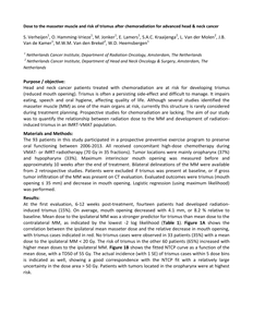
Purpose / objective: Head and neck cancer patients treated with chemoradiation are at risk for developing trismus (reduced mouth opening). Trismus is often a persisting side-effect and difficult to manage. It impairs eating, speech and oral hygiene, affecting quality of life. Although several studies identified the masseter muscle (MM) as one of the main organs at risk, currently this structure is rarely considered during treatment planning. Prospective studies for chemoradiation are lacking. The aim of our study was to quantify the relationship between radiation dose to the MM and development of radiation-induced trismus in an IMRT-VMAT population. Results: At the first evaluation, 6-12 weeks post-treatment, fourteen patients had developed radiation-induced trismus (15%). On average, mouth opening decreased with 4.1 mm, or 8.2 % relative to baseline. Mean dose to the ipsilateral MM was a stronger predictor for trismus than mean dose to the contralateral MM, as indicated by the lowest -2 log likelihood (Table 1). Figure 1A shows the correlation between the ipsilateral mean masseter dose and the relative decrease in mouth opening, with trismus cases indicated in red. No trismus cases were observed in 33 patients (35%) with a mean dose to the ipsilateral MM < 20 Gy. The risk of trismus in the other 60 patients (65%) increased with higher mean doses to the ipsilateral MM. Figure 1B shows the fitted NTCP curve as a function of the mean dose, with a TD50 of 55 Gy. The actual incidence (with 1 SE) of trismus cases within 5 dose bins is indicated as well, showing a good correspondence with the NTCP fit with a relatively large uncertainty in the dose area > 50 Gy. Patients with tumors located in the oropharynx were at highest risk.
DOCUMENT

Op verzoek van Jelle Scheurleer: Purpose: To investigate the accuracy of dose calculation on cone beam CT (CBCT) data sets after HU-RED calibration and validation in phantom studies and clinical patients. Material and methods: Calibration of HU-RED curves for kV-CBCT were generated for three clinical protocols (H&N, thorax and pelvis) by using a Gammex RMI phantom with human tissue equivalent inserts and additional perspex blocks to account for patient scatter. Two calibration curves per clinical protocol were defined, one for the Varian Truebeam 2.0 and another for the OBI systems (Varian, Palo Ato). Differences in HU values with respect to the CT-calibration curve were evaluated for all the inserts. Four radiotherapy plans (breast, prostate, H&N and lung) were produced on an anthropomorphic phantom (Alderson) to evaluate dose differences on the kV-CBCT with the new calibration curves with respect to the CT based dose calculation. Dose differences were evaluated according to the D2%, D98% and Dmean metrics extracted from the DVHs of the plans and - evaluation (2%, 1mm) on the three planes at the isocenter for all plans. Clinical evaluation was performed on 5 patients and dose differences were evaluated as in the phantom study.
DOCUMENT

Introduction: The purpose of this review is to gather and analyse current research publications to evaluate Sinogram-Affirmed Iterative Reconstruction (SAFIRE). The aim of this review is to investigate whether this algorithm is capable of reducing the dose delivered during CT imaging while maintainingimage quality. Recent research shows that children have a greater risk per unit dose due to increased radiosensitivity and longer life expectancies, which means it is particularly important to reduce the radiation dose received by children.Discussion: Recent publications suggest that SAFIRE is capable of reducing image noise in CT images, thereby enabling the potential to reduce dose. Some publications suggest a decrease in dose, by up to 64% compared to filtered back projection, can be accomplished without a change in image quality.However, literature suggests that using a higher SAFIRE strength may alter the image texture, creating an overly ‘smoothed’ image that lacks contrast. Some literature reports SAFIRE gives decreased low contrast detectability as well as spatial resolution. Publications tend to agree that SAFIRE strength threeis optimal for an acceptable level of visual image quality, but more research is required. The importance of creating a balance between dose reduction and image quality is stressed. In this literature review most of the publications were completed using adults or phantoms, and a distinct lack of literature forpaediatric patients is noted.Conclusion: It is necessary to find an optimal way to balance dose reduction and image quality. More research relating to SAFIRE and paediatric patients is required to fully investigate dose reduction potential in this population, for a range of different SAFIRE strengths.
DOCUMENT

Purpose: To determine whether using different combinations of kVp and mAs with additional filtration can reduce the effective dose to a paediatric phantom whilst maintaining diagnostic image quality.Methods: 27 images of a paediatric AP pelvis phantom were acquired with different kVp, mAs and additional copper filtration. Images were displayed on quality controlled monitors with dimmed lighting. Ten diagnostic radiographers (5 students and 5 experienced radiographers) had eye tests to assess visual acuity before rating the images. Each image was rated for visual image quality against a reference image using 2 alternative forced choice software using a 5-point Likert scale. Physical measures (SNR and CNR) were also taken to assess image quality.Results: Of the 27 images rated, 13 of them were of acceptable image quality and had a dose lower than the image with standard acquisition parameters. Two were produced without filtration, 6 with 0.1mm and 5 with 0.2mm copper filtration. Statistical analysis found that the inter-rater and intra-raterreliability was high.Discussion: It is possible to obtain an image of acceptable image quality with a dose that is lower than published guidelines. There are some areas of the study that could be improved. These include using a wider range of kVp and mAs to give an exact set of parameters to use.Conclusion: Additional filtration has been identified as amajor tool for reducing effective dose whilst maintaining acceptable image quality in a 5 year old phantom.
DOCUMENT

This paper reviews the literature for lowering of dose to paediatric patients through use of exposure factors and additional filtration. Dose reference levels set by The International Commission on Radiological Protection (ICRP) will be considered. Guidance was put in place in 1996 requires updatingto come into line with modern imaging equipment. There is a wide range of literature that specifies that grids should not be used on paediatric patients. Although much of the literature advocates additional filtration, contrasting views on the relative benefits of using aluminium or copper filtration, and theireffects on dose reduction and image quality can vary. Changing kVp and mAs has an effect on the dose to the patient and image quality. Collimation protects adjacent structures whilst reducing scattered radiation.
DOCUMENT

The objective of this study was to introduce a new iterative method to reconstruct multi leaf collimator (MLC) positions based on low resolution ionization detector array measurements and to evaluate its error detection performance. The iterative reconstruction method consists of a fluence model, a detector model and an optimizer. Expected detector response was calculated using a radiotherapy treatment plan in combination with the fluence model and detector model. MLC leaf positions were reconstructed by minimizing differences between expected and measured detector response. The iterative reconstruction method was evaluated for an Elekta SLi with 10.0 mm MLC leafs in combination with the COMPASS system and the MatriXX Evolution (IBA Dosimetry) detector with a spacing of 7.62 mm. The detector was positioned in such a way that each leaf pair of the MLC was aligned with one row of ionization chambers. Known leaf displacements were introduced in various field geometries ranging from −10.0 mm to 10.0 mm. Error detection performance was tested for MLC leaf position dependency relative to the detector position, gantry angle dependency, monitor unit dependency, and for ten clinical intensity modulated radiotherapy (IMRT) treatment beams. For one clinical head and neck IMRT treatment beam, influence of the iterative reconstruction method on existing 3D dose reconstruction artifacts was evaluated. The described iterative reconstruction method was capable of individual MLC leaf position reconstruction with millimeter accuracy, independent of the relative detector position within the range of clinically applied MU's for IMRT. Dose reconstruction artifacts in a clinical IMRT treatment beam were considerably reduced as compared to the current dose verification procedure. The iterative reconstruction method allows high accuracy 3D dose verification by including actual MLC leaf positions reconstructed from low resolution 2D measurements.
LINK
Objective: Summarize all relevant findings in published literature regarding the potential dose reduction related to image quality using Sinogram-Affirmed Iterative Reconstruction (SAFIRE) compared to Filtered Back Projection (FBP).Background: Computed Tomography (CT) is one of the most used radiographic modalities in clinical practice providing high spatial and contrast resolution. However it also delivers a relatively high radiation dose to the patient. Reconstructing raw-data using Iterative Reconstruction (IR) algorithmshas the potential to iteratively reduce image noise while maintaining or improving image quality of low dose standard FBP reconstructions. Nevertheless, long reconstruction times made IR unpractical for clinical use until recently.Siemens Medical developed a new IR algorithm called SAFIRE, which uses up to 5 different strength levels, and poses an alternative to the conventional IR with a significant reconstruction time reduction.Methods: MEDLINE, ScienceDirect and CINAHL databases were used for gathering literature. Eleven articles were included in this review (from 2012 to July 2014).Discussion: This narrative review summarizes the results of eleven articles (using studies on both patients and phantoms) and describes SAFIRE strengths for noise reduction in low dose acquisitions while providing acceptable image quality.Conclusion: Even though the results differ slightly, the literature gathered for this review suggests that the dose in current CT protocols can be reduced at least 50% while maintaining or improving image quality. There is however a lack of literature concerning paediatric population (with increased radiationsensitivity). Further studies should also assess the impact of SAFIRE on diagnostic accuracy.
DOCUMENT
