Medical imaging practice changed dramatically with the introduction of digital imaging. Although digital imaging has many advantages, it also has made it easier to delete images that are not of diagnostic quality. Mistakes in imaging—from improper patient positioning, patient movement during the examination, and selecting improper equipment—could go undetected when images are deleted. Such an approach would preclude a reject analysis from which valuable lessons could be learned. In the analog days of radiography, saving the rejected films and then analyzing them was common practice among radiographers. In principle, reject analysis can be carried out easier and with better tools (ie, software) in the digital era, provided that rejected images are stored for analysis. Reject analysis and the subsequent lessons learned could reduce the number of repeat images, thus reducing imaging costs and decreasing patient exposure to radiation. The purpose of this study, which was conducted by order of the Dutch Healthcare Inspectorate, was to investigate whether hospitals in the Netherlands store and analyze failed imaging and, if so, to identify the tools used to analyze those images.
DOCUMENT

Review: With great interest we have read the paper “Pregnancy Screening before Diagnostic Radiography in Emergency Department; an Educational Review” by A.I. Abushouk et al. (1). We agree with the authors that unnecessary fetal radiation exposure should be avoided and that pregnancy screening can be a means to accomplish this. However, in their paper the authors suggest in several instances that radiological imaging during pregnancy can lead to teratogenic effects. In the Abstract it is stated: “Radiation exposure during pregnancy may have serious teratogenic effects to the fetus. Therefore, checking the pregnancy status before imaging women of child bearing age can protect against these effects.”, and in the Introduction: “Therefore, checking the pregnancy status before imaging women of child bearing age can protect against radiation teratogenic effects.” We strongly disagree with these statements: common radiological imaging will usually not give rise to fetal radiation doses high enough to lead to teratogenesis. The statements in the paper may lead to unnecessary worrying of pregnant women and it may discourage themfrom undergoing medically necessary radiological examinations.
DOCUMENT

Artificial Intelligence (AI) has changed radiology substantially in the last years, where the focus of attention has mainly been on the radiologist. However, the radiographer’s role has been largely ignored even though AI is also affecting for example patient positioning, treatment planning and image reconstruction: tasks that are typically carried out by radiographers (and RTTs). Radiographers are currently not prepared for the changes in their profession that will come with the introduction of AI into everyday work.
DOCUMENT

Abstract gepubliceerd in Elsevier: Introduction: Recent research has identified the issue of ‘dose creep’ in diagnostic radiography and claims it is due to the introduction of CR and DR technology. More recently radiographers have reported that they do not regularly manipulate exposure factors for different sized patients and rely on pre-set exposures. The aim of the study was to identify any variation in knowledge and radiographic practice across Europe when imaging the chest, abdomen and pelvis using digital imaging. Methods: A random selection of 50% of educational institutes (n ¼ 17) which were affiliated members of the European Federation of Radiographer Societies (EFRS) were contacted via their contact details supplied on the EFRS website. Each of these institutes identified appropriate radiographic staff in their clinical network to complete an online survey via SurveyMonkey. Data was collected on exposures used for 3 common x-ray examinations using CR/DR, range of equipment in use, staff educational training and awareness of DRL. Descriptive statistics were performed with the aid of Excel and SPSS version 21. Results: A response rate of 70% was achieved from the affiliated educational members of EFRS and a rate of 55% from the individual hospitals in 12 countries across Europe. Variation was identified in practice when imaging the chest, abdomen and pelvis using both CR and DR digital systems. There is wide variation in radiographer training/education across countries.
DOCUMENT

Introduction: In the Netherlands, Diagnostic Reference Levels (DRLs) have not been based on a national survey as proposed by ICRP. Instead, local exposure data, expert judgment and the international scientific literature were used as sources. This study investigated whether the current DRLs are reasonable for Dutch radiological practice. Methods: A national project was set up, in which radiography students carried out dose measurements in hospitals supervised by medical physicists. The project ran from 2014 to 2017 and dose values were analysed for a trend over time. In the absence of such a trend, the joint yearly data sets were considered a single data set and were analysed together. In this way the national project mimicked a national survey. Results: For six out of eleven radiological procedures enough data was collected for further analysis. In the first step of the analysis no trend was found over time for any of these procedures. In the second step the joint analysis lead to suggestions for five new DRL values that are far below the current ones. The new DRLs are based on the 75 percentile values of the distributions of all dose data per procedure. Conclusion: The results show that the current DRLs are too high for five of the six procedures that have been analysed. For the other five procedures more data needs to be collected. Moreover, the mean weights of the patients are higher than expected. This introduces bias when these are not recorded and the mean weight is assumed to be 77 kg. Implications for practice: The current checking of doses for compliance with the DRLs needs to be changed. Both the procedure (regarding weights) and the values of the DRLs should be updated.
MULTIFILE

Chest imaging plays a pivotal role in screening and monitoring patients, and various predictive artificial intelligence (AI) models have been developed in support of this. However, little is known about the effect of decreasing the radiation dose and, thus, image quality on AI performance. This study aims to design a low-dose simulation and evaluate the effect of this simulation on the performance of CNNs in plain chest radiography. Seven pathology labels and corresponding images from Medical Information Mart for Intensive Care datasets were used to train AI models at two spatial resolutions. These 14 models were tested using the original images, 50% and 75% low-dose simulations. We compared the area under the receiver operator characteristic (AUROC) of the original images and both simulations using DeLong testing. The average absolute change in AUROC related to simulated dose reduction for both resolutions was <0.005, and none exceeded a change of 0.014. Of the 28 test sets, 6 were significantly different. An assessment of predictions, performed through the splitting of the data by gender and patient positioning, showed a similar trend. The effect of simulated dose reductions on CNN performance, although significant in 6 of 28 cases, has minimal clinical impact. The effect of patient positioning exceeds that of dose reduction.
LINK
Dit artikel is een vertaling van het artikel “Digital Radiography Reject Analysis: Results of a Survey Among Dutch Hospitals” dat in de mei/juni 2020 editie van het blad Radiologic Technology is gepubliceerd. Korte samenvatting: In opdracht van de Inspectie voor de Gezondheidszorg is aan een steekproef van Nederlandse ziekenhuizen gevraagd hoe zij omgaan met medische beelden die worden afgekeurd. De resultaten laten zien dat de meeste ziekenhuizen deze opnames niet bewaren voor analyse.
DOCUMENT

Introduction: Zygomatic fractures can be diagnosed with either computed tomography (CT) or direct digital radiography (DR). The aim of the present study was to assess the effect of CT dose reduction on the preference for facial CT versus DR for accurate diagnosis of isolated zygomatic fractures. Materials and methods: Eight zygomatic fractures were inflicted on four human cadavers with a free fall impactor technique. The cadavers were scanned using eight CT protocols, which were identical except for a systematic decrease in radiation dose per protocol, and one DR protocol. Single axial CT images were displayed alongside a DR image of the same fracture creating a total of 64 dual images for comparison. A total of 54 observers, including radiologists, radiographers and oral and maxillofacial surgeons, made a forced choice for either CT or DR. Results: Forty out of 54 observers (74%) preferred CT over DR (all with P < 0.05). Preference for CT was maintained even when radiation dose reduced from 147.4 mSv to 46.4 mSv (DR dose was 6.9 mSv). Only a single out of all raters preferred DR (P ¼ 0.0003). The remaining 13 observers had no significant preference. Conclusion: This study demonstrates that preference for axial CT over DR is not affected by substantial (~70%) CT dose reduction for the assessment of zygomatico-orbital fractures.
MULTIFILE

This review aims to identify strategies to optimise radiography practice using digital technologies, for full spine studies on paediatrics focusing particularly on methods used to diagnose and measure severity of spinal curvatures. The literature search was performed on different databases (PubMed, Google Scholar and ScienceDirect) and relevant websites (e.g., American College of Radiology and International Commission on Radiological Protection) to identify guidelines and recent studies focused on dose optimisation in paediatrics using digital technologies. Plain radiography was identified as the most accurate method. The American College of Radiology (ACR) and European Commission (EC) provided two guidelines that were identified as the most relevant to the subject. The ACR guidelines were updated in 2014; however these guidelines do not provide detailed guidance on technical exposure parameters. The EC guidelines are more complete but are dedicated to screen film systems. Other studies provided reviews on the several exposure parameters that should be included for optimisation, such as tube current, tube voltage and source-to-image distance; however, only explored few of these parameters and not all of them together. One publication explored all parameters together but this was for adults only. Due to lack of literature on exposure parameters for paediatrics, more research is required to guide and harmonise practice
DOCUMENT

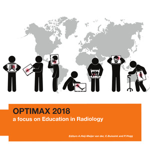
This year, OPTIMAX was warmly welcomed by University College Dublin. For the sixth time students and teachers from Europe, South Africa, South America and Canada have come together enthusiastically to do research in the Radiography domain. As in previous years, there were several research groups consisting of PhD-, MSc- and BSc students and tutors from the OPTIMAX partner Universities or on invitation by partner Universities. OPTIMAX 2018 was partly funded by the partner Universities and partly by the participants.
DOCUMENT
