DOCUMENT
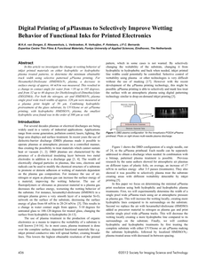
Light scattering is a fundamental property that can be exploited to create essential devices such as particle analysers. The most common particle size analyser relies on measuring the angle-dependent diffracted light from a sample illuminated by a laser beam. Compared to other non-light-based counterparts, such a laser diffraction scheme offers precision, but it does so at the expense of size, complexity and cost. In this paper, we introduce the concept of a new particle size analyser in a collimated beam configuration using a consumer electronic camera and machine learning. The key novelty is a small form factor angular spatial filter that allows for the collection of light scattered by the particles up to predefined discrete angles. The filter is combined with a light-emitting diode and a complementary metal-oxide-semiconductor image sensor array to acquire angularly resolved scattering images. From these images, a machine learning model predicts the volume median diameter of the particles. To validate the proposed device, glass beads with diameters ranging from 13 to 125 µm were measured in suspension at several concentrations. We were able to correct for multiple scattering effects and predict the particle size with mean absolute percentage errors of 5.09% and 2.5% for the cases without and with concentration as an input parameter, respectively. When only spherical particles were analysed, the former error was significantly reduced (0.72%). Given that it is compact (on the order of ten cm) and built with low-cost consumer electronics, the newly designed particle size analyser has significant potential for use outside a standard laboratory, for example, in online and in-line industrial process monitoring.
MULTIFILE
Muscle fiber-type specific expression of UCP3-protein is reported here for the firts time, using immunofluorescence microscopy
DOCUMENT

Inkjet printing is a rapidly growing technology for depositing functional materials in the production of organic electronics. Challenges lie among others in the printing of high resolution patterns with high aspect ratio of functional materials to obtain the needed functionality like e.g. conductivity. μPlasma printing is a technology which combines atmospheric plasma treatment with the versatility of digital on demand printing technology to selectively change the wetting behaviour of materials. In earlier research it was shown that with μPlasma printing it is possible to selectively improve the wetting behaviour of functional inks on polymer substrates using atmospheric air plasma. In this investigation we show it is possible to selectively change the substrate wetting behaviour using combinations of different plasmas and patterned printing. For air and nitrogen plasmas, increased wetting of printed materials could be achieved on both polycarbonate and glass substrates. A minimal track width of 320 μm for a 200 μm wide plasma needle was achieved. A combination of N2 with HMDSO plasma increases the contact angle for water up from <100 to 1050 and from 320 to 460 for DEGDMA making the substrate more hydrophobic. Furthermore using N2-plasma in combination with a N2/HMDSO plasma, hydrophobic tracks could be printed with similar minimal track width. Combining both N2 -plasma and N2/HMDSO plasma treatments show promising results to further decrease the track width to even smaller values.
DOCUMENT

In het kader van actualisering van voorlichtingspublicaties (een samenwerkingsverband tussen FDP, FME, NIL, NIMR, Syntens en TNO Industrie & Techniek), is deze voorlichtingspublicatie aangepast aan de huidige stand der techniek. De originele publicatie is in 1992 tot stand gekomen door samenwerking van de Vereniging FME/CWM en het Nederlands Instituut voor Lastechniek in het kader van het FME/NIL project "Het lijmen als verbindingstechniek".
DOCUMENT

Om inzicht te krijgen in spierveroudering is genexpressie gemeten in vastus lateralis biopten van jonge en oude mannen en vrouwen. We vonden dat tijdens het ouder worden bij beide geslachten dezelfde categorieën genen in spieren worden aan- en uitgeschakeld (“gereguleerd”); de mate van deze zogenaamde differentiële expressie was echter geslachtsspecifiek. Bij mannen was oxidatieve fosforylering het meest in het oog springende proces, en bij vrouwen was dit celgroei gemedieerd door AKT-signalering. De conclusie is dat dezelfde processen zijn geassocieerd met skeletspierveroudering bij mannen en vrouwen, maar dat de differentiële expressie van die processen geslachtsspecifiek is.
MULTIFILE

In this article we investigate the change in wetting behavior of inkjet printed materials on either hydrophilic or hydrophobic plasma treated patterns, to determine the minimum obtainable track width using selective patterned μPlasma printing. For Hexamethyl-Disiloxane (HMDSO)/N2 plasma, a decrease in surface energy of approx. 44 mN/m was measured. This resulted in a change in contact angle for water from <10 up to 105 degrees, and from 32 up to 46 degrees for Diethyleneglycol-Dimethaclylate (DEGDMA). For both the nitrogen, air and HMDSO/N2 plasma single pixel wide track widths of approx. 320 μm were measured at a plasma print height of 50 μm. Combining hydrophilic pretreatment of the glass substrate, by UV/Ozone or air μPlasma printing, with hydrophobic HMDSO/N2 plasma, the smallest hydrophilic area found was in the order of 300 μm as well.
DOCUMENT
In manufacturing of organic electronics, inkjet printing as an alternative technique for depositing materials is becoming increasingly important. Aside to the ink formulations challenges, improving the resolution of the printed patterns is a major goal. In this study we will discuss a newly developed technique to selectively modify the substrate surface energy using plasma treatment as a means to achieve this goal. First, we look at the effects of the μPlasma treatment on the surface energy for a selection of plastic films. Second, we investigated the effects of the μPlasma treatment on the wetting behaviour of inkjet printed droplets to determine the resolution of the μPlasma printing technique. We found that the surface energy for all tested films increased significantly reaching a maximum after 3-5 repetitions. Subsequently the surface energy decreased in the following 8-10 days after treatment, finally stabilizing at a surface energy roughly halfway between the surface energy of the untreated film and the maximum obtained surface energy. When μPlasma printing lines, an improved wetting abillity of inkjet printed materials on the plasma treated areas was found. The minimal achieved μPlasma printed line was found to be 1 mm wide. For future application it is important to increase the resolution of the plasma print process. This is crucial for combining plasma treatment with inkjet print technology as a means to obtain higher print resolutions.
DOCUMENT

Deze publicatie is gemaakt om een overzicht te geven van de mogelijkheden van het toepassen van thermisch gespoten aluminium deklagen. Naast deze publicatie verschijnen in het kader van het project "Nieuwe coatingtechnieken voor het MKB" nog vier andere publicaties die gezamenlijk een, zij het niet volledig, beeld geven van coatingtechnologie in het algemeen en de vele aspecten die daarmee samenhangen.
DOCUMENT

Bespreking van onderzoek van Rychard Bouwens in ‘Waar wij trots op zijn. De ontdekkingen van 2011’ van de Universiteit Leiden Faculteit der Wiskunde & Natuurwetenschappen. Het valt goed te begrijpen voor iedereen met een basale kennis van klassieke fotografie: bij weinig licht neem je een lange sluitertijd. En dat is wat Rychard Bouwens deed. Om naar de zogenaamde Dark Ages van het heelal te kijken, hield hij de Hubble-ruimtetelescoop maar liefst 87 uur lang op een plek gericht.
DOCUMENT

